Recurring Brain Tumor vs Mommy of Two with Gamma Knife
Round I consisted of a crainiotomy to remove the newly discovered,
hemorrhaging brain tumor the size of a baseball. I was 9m pregnant
with my now 3 1/2 year old daughter. The day after she was born by emergency
c-section, I underwent a 14 hour surgery with two neurosurgeons. They cut my
scalp from ear to ear, peeled it back, and drilled six bore holes on the top left of my head, sawed between the holes, and set the piece aside. Then they advanced on
the highly vascular tumor lurking deep within my brain, in and around the left lateral ventrical (there are 4 of these cerebral spinal fluid filled spaces). It required hours upon hours of highly detailed work, including cauterizing thousands of vessels.
Eventually, they got it all out and stapled my head back together. Three long weeks later, I finally went home, only to return after a week for a second surgery.
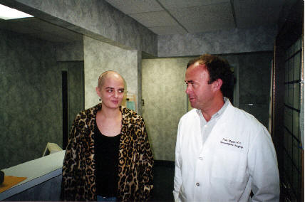
Me and my doctor
I had a trapped ventricle due to scarring, so they had to install a ventricular/peritoneal shunt
with a tube from the ventricle coming out the back of my head, and down around to my abdominal cavity, all underneath the skin. They did part of it laproscopically, so I had a tiny incision below my belly button as well. The shunt tube looks like a big vein, if you happen to see it.
I have had many MRI's and CAT scans over the last few years. Cat scans are relativly simple, but the MRI sounds kind of like a weird sort of jackhammer. Very loud! Every September, I have a follow-up MRI to check for regrowth, as I didn't have radiation or chemo previously. The 2000 scan actually showed a 1.5 cm growth, but it was not compared with the '99 scan, since I had it done at another MRI center and my former neurosurgeon wasn't taking medicaid anymore. So last November, after my son was born in October, I had my annual MRI two months late. My opponent was back for more.
As it was relatively small yet at about 2.5cm, I was a candidate for Ft. Worth's new (December 2001) Gamma Knife instead of going through all that again. It seems to have gone quite well, I'm feeling only slightly bad. Also, requested and got morphine after I was done with the MRI part.
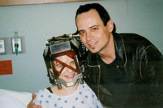
In my GK MRI brain mapping helmet with sweetheart
The pressure of the frame was unbearable. It felt like it was crushing my skull. I am now taking VicodenES twice a day. I only have 4 small holes in my head that hurt some, but they're nothing compared to a massive incision, and hair loss. I have 2 band aids on my forehead. No problem. The tumor will now become necrotic and shrivel up. Hear me tumor? Or else!
Here' s a couple exerpts from my neurosurgeons website (www.cndpa.com):
 When you first arrive to the unit, you will change into a hospital gown, the nurse will start an IV, you will be attached to an EKG monitor, and the nurse will administer medications to help you relax and for prevention of nausea. Your procedure will begin with placement of an aluminum head frame, which is held into place by 4 screw-like pins in the very outer layer of the skull. You will have 2 pins inserted to your forehead and 2 pins inserted to the back of your head. Prior to the insertion of the head frame, numbing cream will be applied to the 4 pin site areas and then a local anesthetic will be administered to each pin site area. You may feel some discomfort for a short time. Additional medication to help you relax during head frame placement can be given if needed. When you first arrive to the unit, you will change into a hospital gown, the nurse will start an IV, you will be attached to an EKG monitor, and the nurse will administer medications to help you relax and for prevention of nausea. Your procedure will begin with placement of an aluminum head frame, which is held into place by 4 screw-like pins in the very outer layer of the skull. You will have 2 pins inserted to your forehead and 2 pins inserted to the back of your head. Prior to the insertion of the head frame, numbing cream will be applied to the 4 pin site areas and then a local anesthetic will be administered to each pin site area. You may feel some discomfort for a short time. Additional medication to help you relax during head frame placement can be given if needed.
After placement of the head frame, you will be taken to radiology where an MRI will be obtained for localizing and planning your Gamma Knife treatment. After completion of your MRI, you will return to the Gamma Knife unit where you will stay while the physicians and physicist study your films and "map" out your treatment time. This may take 1-2 hours. During this time you may watch television and visit with family members.
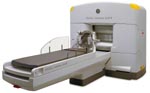
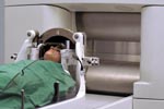 When treatment planning is completed, you will be moved into the Gamma Knife treatment room where you will be placed on the "couch" or Gamma Knife table. A large silver helmet will be attached to the table, and you head frame will be attached to this. Once all of the coordinates are set for your treatment, a microphone will be attached to your hospital gown, and the Gamma Knife team will leave the room. Monitors are set up for the physicians and physicist to watch you during the procedure, and the microphone will allow you to communicate with them. When the procedure begins, part of the Gamma Knife table will slide back into the dome, and the actual radiation treatment will begin. Your treatment may last anywhere from 30 minutes to 2 hours depending on you diagnosis. When treatment planning is completed, you will be moved into the Gamma Knife treatment room where you will be placed on the "couch" or Gamma Knife table. A large silver helmet will be attached to the table, and you head frame will be attached to this. Once all of the coordinates are set for your treatment, a microphone will be attached to your hospital gown, and the Gamma Knife team will leave the room. Monitors are set up for the physicians and physicist to watch you during the procedure, and the microphone will allow you to communicate with them. When the procedure begins, part of the Gamma Knife table will slide back into the dome, and the actual radiation treatment will begin. Your treatment may last anywhere from 30 minutes to 2 hours depending on you diagnosis.
After your treatment is completed, you will be removed from the Gamma Knife table, returned to the frame room and the head frame will be removed. A band-aid will be applied to each pin site. At this time you may have something to drink and a light snack. Your total time in the Gamma Knife unit will be 4-5 hours. Approximately 30-minutes to 1 hour after your treatment completes, you will be transferred to your hospital room for an overnight stay for observation.
Gamma Knife Radiosurgery
Revolutionary Noninvasive Brain Surgery
At 20 tons, this is not a knife in any usual sense. Actually, the Gamma Knife® is a device for a form of neurosurgery that requires no incision. It can reduce surgical complications and shortens the average patient's hospital stay to an outpatient procedure.
How it Works
The Gamma Knife® directs 201 beams of ionizing gamma radiation to a targeted area within the brain. Intersecting at the target, the concentrated rays attempt to eradicate small- to medium-sized tumors and vascular abnormalities. Meanwhile, surrounding tissues receive minimal radiation exposure. Highly sophisticated, three-dimensional computer guidance gives the Gamma Knife its amazing exactness--accurate down to one half of a millimeter, about the width of a human hair.Developed by Swedish Professor Lars Leksell, this unique neurosurgical tool combines a stereotactic frame (for head support and radiation targeting) with a collimator helmet (for radiation guidance) and a source of radiation (Cobalt-60).
Potential Candidates for Gamma Knife Radiosurgery
- Patients with benign or malignant intracranial tumors, vascular malformation, or trigeminal neuralgia.
- Patients with surgically inaccessible brain lesions.
- Patients who are medically unable to undergo conventional surgery.
- Patients with recurrent tumors.
- Patients whose tumors could not be completely removed with conventional surgery
- Patients of all ages, including children.
Advantages of Gamma Knife Treatment
The Gamma Knife ensures only abnormal tissue is treated while surrounding healthy tissue remains unaffected.
Because the Gamma Knife does not require an incision, risk associated with infection and bleeding is not a concern.
While conventional neurosurgery requires lengthy hospital stays and a potentially long recovery period, Gamma Knife patients are able to resume normal activities within 24-48 hours.
Gamma Knife radiosurgery costs 30-70% less than a craniotomy.
Gamma Knife radiosurgery is a one time treatment. (I hope!)
after the first surgery,
my entire right side was paralyzed, and
I also had severe edema in my head because I
pulled one of the drainage tubes out.
I looked like the great pumpkinhead for a few days (luckily there are no pictures of that)!
I also had Aphasia, which for me meant trouble with verbal expression.
Eventually I have regained those abilities.
With the Gamma Knife, I have been able to avoid all such problems.
A technological wonder! =)


|